
Post Category


You may have heard "macular degeneration" used as if it were a single diagnosis with a single outlook. It is not. Age-related macular degeneration (AMD) is one condition with two very different forms, and the distinction matters enormously, because they progress at different speeds and are treated in completely different ways. The dry form is far more common and usually advances slowly over years. The wet form is less common but can steal central vision in a matter of weeks if it is not treated promptly. Understanding which form you have, and recognizing the warning signs that dry can convert to wet, is one of the most important things a patient over 60 can do for their sight.
AMD is the leading cause of severe central vision loss in adults over 60 in the developed world, and South Florida has one of the oldest patient populations in the country. In Boca Raton, this is a conversation we have nearly every day. This guide explains what the macula is, how the two forms differ, how AMD is diagnosed and monitored, and what the 2026 treatment landscape actually offers, including newer injections for geographic atrophy. It is written for an educated patient and is not a substitute for a dilated eye examination.
Macular degeneration is one diagnosis with two very different paths
The single most useful thing to understand about AMD is that "wet" and "dry" are not two separate diseases. They are two stages and forms of the same underlying condition. Almost everyone with AMD starts with the dry form. A minority of those patients go on to develop the wet form, which is why ongoing monitoring is so important even when the disease seems stable. The dry form is managed with nutrition, monitoring, and, in advanced cases, newer therapies. The wet form is a same-week problem treated with injections. Mistaking one for the other, or assuming "dry and stable" means "no need to watch," is the error that costs people vision.
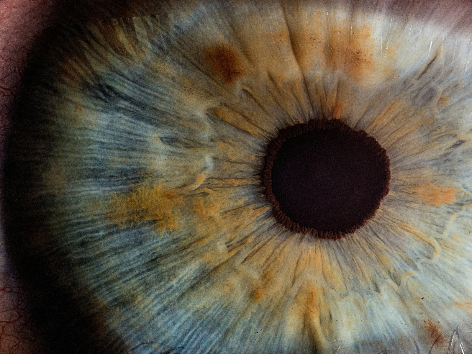
What macular degeneration actually is
The macula is the small, central part of the retina responsible for your sharpest, most detailed vision: reading, recognizing faces, seeing fine print, and driving. AMD is the breakdown of this central area, while the peripheral retina, and therefore your side vision, is typically spared. That is why people with advanced AMD rarely go completely blind, but they can lose the detailed central vision that most daily tasks depend on.
According to the National Eye Institute, AMD is driven by aging changes in the macula, with genetics, smoking, cardiovascular health, and lifetime sun exposure all contributing to risk. It does not cause pain, and early on it often causes no symptoms at all, which is exactly why it is so frequently caught first on a routine dilated exam.
Dry macular degeneration
Dry AMD (also called non-neovascular or atrophic AMD) accounts for the large majority of cases. It develops as the macula accumulates small yellow deposits called drusen and as the supporting cells of the retina gradually thin and break down. Dry AMD is usually staged in three steps:
- Early dry AMD. Small to medium drusen are present, usually with no noticeable vision changes.
- Intermediate dry AMD. Larger drusen and pigment changes appear. Some patients begin to notice mild blur or difficulty in low light. This is the stage where nutritional supplements become relevant.
- Advanced dry AMD (geographic atrophy). Areas of retinal cells die off in well-defined patches called geographic atrophy (GA). As these patches enlarge and reach the center of the macula, central vision is lost. GA tends to progress slowly but relentlessly.
Dry AMD usually progresses over years, not weeks. The main risks are gradual loss of central detail and, critically, the possibility of converting to the wet form. That is why monitoring never stops.
Wet macular degeneration
Wet AMD (also called neovascular AMD) is less common but far more aggressive. It occurs when abnormal new blood vessels grow beneath the retina, a process called choroidal neovascularization. These vessels are fragile and leak fluid and blood into and under the macula. That fluid distorts and damages the delicate central retina, and it can do so quickly.
The hallmark symptom is the relatively sudden onset of distortion: straight lines (a door frame, a window blind, lines of text) begin to look wavy or bent, a blurry or dark spot appears in the center of vision, or vision drops noticeably over days to weeks. Wet AMD is the form responsible for most cases of rapid, severe central vision loss from AMD. The good news is that it is treatable, often very effectively, when caught early, which is why these symptoms are a same-week emergency, not something to watch and wait on.
How macular degeneration is diagnosed
Diagnosis and monitoring rely on several tools, used together:
- Dilated eye exam. The foundation. With the pupils dilated, your ophthalmologist examines the macula directly for drusen, pigment changes, atrophy, fluid, or bleeding.
- Optical coherence tomography (OCT). A non-invasive scan that produces a cross-section of the retina, showing fluid, thickness changes, and atrophy with great precision. OCT is the workhorse for detecting wet AMD activity and tracking response to treatment.
- Fluorescein or OCT angiography. Imaging of the retinal and choroidal blood vessels to identify and map abnormal leaking vessels in suspected wet AMD.
- Amsler grid. A simple home monitoring tool, a grid of straight lines with a central dot. Patients check each eye separately, and any new waviness, distortion, or missing area is reported to their ophthalmologist promptly. The American Academy of Ophthalmology recommends regular home Amsler grid testing for many patients with AMD.
Because early and intermediate dry AMD often have no symptoms, regular dilated exams are the only reliable way to catch AMD before it advances. This is a core reason we emphasize the comprehensive eye exam for older adults, even those who feel their vision is fine.
Treating dry AMD in 2026
For most of the history of AMD care, there was no treatment for the dry form, only monitoring. That has changed.
AREDS2 supplements. For patients with intermediate or advanced AMD in at least one eye, a specific formulation of vitamins and minerals can reduce the risk of progression to advanced disease. The AREDS2 trial run by the National Eye Institute established the modern formula (vitamin C, vitamin E, lutein, zeaxanthin, zinc, and copper). AREDS2 supplements do not cure AMD or restore lost vision, and they are not recommended for patients with only early AMD or no AMD. They reduce the risk of progression in the right candidates, which is a meaningful but specific benefit your ophthalmologist will assess for you.
Lifestyle. Not smoking, controlling blood pressure and cardiovascular risk, eating a diet rich in leafy greens and fish, and protecting the eyes from ultraviolet light are all associated with lower AMD risk and progression. In South Florida's year-round sun, UV-blocking sunglasses are an easy, sensible measure.
Complement-inhibitor injections for geographic atrophy. A genuinely new development is the availability of injectable treatments for geographic atrophy, the advanced form of dry AMD. The FDA has approved complement-inhibitor injections, pegcetacoplan and avacincaptad pegol, that have been shown to slow the growth of geographic atrophy lesions over time. It is important to be clear about what these do and do not do: they are shown to slow the rate at which GA expands, but they do not reverse atrophy or restore vision that is already lost, and they require ongoing injections with their own risks. Whether they are appropriate is an individualized decision made with a retina specialist.
Vision rehabilitation. For vision already lost to advanced AMD, low-vision rehabilitation, magnifiers, and adaptive technology help patients keep reading, recognizing faces, and living independently.
Treating wet AMD in 2026
The treatment of wet AMD is one of the genuine success stories of modern ophthalmology. The mainstay is anti-VEGF therapy.
How anti-VEGF works. The abnormal vessels in wet AMD are driven by a signaling protein called vascular endothelial growth factor (VEGF). Anti-VEGF medications block that signal, which causes the leaking vessels to regress and the macular fluid to dry up. Multiple anti-VEGF agents are available in 2026, and the choice of agent and dosing schedule is a clinical decision made by your retina specialist based on your eye's response. We do not name a single "best" drug, because the right one depends on the individual.
What an injection visit looks like. The eye is numbed thoroughly with anesthetic, then cleaned with an antiseptic. The medication is delivered through a very fine needle into the vitreous (the gel inside the eye). The injection itself takes only seconds, and most patients are surprised by how tolerable it is. You will be monitored briefly and sent home with instructions.
The schedule. Treatment usually begins with a series of monthly injections, after which many patients move to an extended-interval approach (often described as "treat and extend"), where the interval between injections is gradually lengthened as long as the macula stays dry. Some patients need ongoing injections indefinitely. Consistency matters: skipped treatments can allow the disease to reactivate and cause irreversible damage.
Outcomes. When started promptly, anti-VEGF therapy stabilizes vision in the large majority of wet AMD patients and improves vision in a meaningful share of them. Outcomes are best when treatment begins before significant scarring has occurred, which again is why new distortion is a same-week problem. The American Society of Retina Specialists provides patient resources on what to expect from anti-VEGF therapy. Treatment does not cure the underlying AMD, and it is not risk-free, but for a condition that once meant near-certain central vision loss, it is transformative.
When to call your ophthalmologist immediately
Contact your ophthalmologist right away, do not wait for your next routine appointment, if you notice any of the following in either eye:
- Straight lines that suddenly appear wavy, bent, or distorted.
- A new blurry, dark, or empty spot in your central vision.
- A noticeable drop in central vision over days.
- New difficulty reading or recognizing faces that came on quickly.
- Any change on your home Amsler grid.
These can be signs that dry AMD has converted to wet AMD, where time directly affects how much vision can be saved.
Living with AMD in South Florida
For our older Boca Raton and Palm Beach County patients, a few local realities are worth naming. Year-round ultraviolet exposure means UV-blocking sunglasses and a hat are sensible daily habits, not just beach-day items. Driving independence is often a central concern, and low-vision tools combined with timely treatment help many patients keep driving safely longer; your ophthalmologist and the Florida licensing standards together guide that decision. And because so much daily life here is outdoors and social, vision rehabilitation services that preserve reading and face recognition have an outsized quality-of-life impact. AMD is also frequently managed alongside other age-related eye conditions, so coordinated retina care matters. Patients with diabetes have additional reasons for close monitoring, since diabetic retinopathy can coexist with AMD.
Important Safety Information
This article is for general education and is not medical advice. AMD treatment decisions, including AREDS2 supplementation, complement-inhibitor injections for geographic atrophy, and anti-VEGF therapy for wet AMD, must be individualized by a qualified ophthalmologist or retina specialist. AREDS2 supplements are intended for specific stages of AMD and are not appropriate for everyone; discuss them with your physician before starting, particularly if you smoke or have other health conditions. Intravitreal injections (anti-VEGF and complement inhibitors) carry risks that include, but are not limited to, eye infection (endophthalmitis), retinal detachment, intraocular pressure elevation, intraocular inflammation, and, rarely, serious vision loss. Complement-inhibitor therapy for geographic atrophy slows lesion growth but does not restore lost vision and has been associated with a risk of conversion to wet AMD in some patients. Wet AMD is urgent but is not universally treatable or risk-free, and outcomes depend on how early treatment begins. Any sudden change in central or distorted vision requires prompt evaluation by an ophthalmologist.
Frequently Asked Questions
What is the difference between wet and dry macular degeneration?
Both are forms of age-related macular degeneration. Dry AMD is far more common, develops slowly over years as the macula thins and accumulates deposits called drusen, and in its advanced stage causes geographic atrophy. Wet AMD is less common but more aggressive, caused by abnormal leaking blood vessels under the retina, and can cause rapid central vision loss within weeks if untreated.
Can dry macular degeneration turn into wet?
Yes. A minority of patients with dry AMD go on to develop the wet form, sometimes suddenly. This is why ongoing monitoring with dilated exams and a home Amsler grid is so important, and why any new distortion or central vision change should be reported to your ophthalmologist immediately.
Do AREDS2 supplements cure macular degeneration?
No. AREDS2 supplements do not cure AMD or restore lost vision. In patients with intermediate or advanced AMD in at least one eye, the AREDS2 formula has been shown to reduce the risk of progression to advanced disease. They are not recommended for people with only early AMD or no AMD. Your ophthalmologist will determine whether they are right for you.
How do anti-VEGF injections for wet AMD work?
Wet AMD is driven by a protein called VEGF that fuels abnormal, leaking blood vessels under the retina. Anti-VEGF medications, injected into the eye, block that signal so the vessels regress and the macular fluid clears. Treatment typically begins monthly and is then extended as the macula stays dry. The injection takes only seconds and is performed after the eye is numbed.
Are there new treatments for advanced dry AMD?
Yes. The FDA has approved complement-inhibitor injections (pegcetacoplan and avacincaptad pegol) that slow the growth of geographic atrophy, the advanced form of dry AMD. They slow the progression of atrophy but do not reverse damage or restore lost vision, and they carry their own risks. Whether they are appropriate is an individualized decision with a retina specialist.
What is an Amsler grid and how do I use it?
An Amsler grid is a simple square grid of straight lines with a dot in the center. You cover one eye, focus on the central dot, and check whether any lines look wavy, blurry, or missing, then repeat with the other eye. Any new distortion is a reason to contact your ophthalmologist promptly, because it can signal a conversion to wet AMD.
Will I go blind from macular degeneration?
AMD affects central vision and usually spares peripheral (side) vision, so it rarely causes total blindness. However, advanced AMD can cause significant loss of the detailed central vision needed for reading, driving, and recognizing faces. Timely treatment, monitoring, and low-vision rehabilitation help many patients preserve function and independence.
How often should I have my eyes checked if I have AMD?
The interval depends on the stage and form of your AMD and is set by your ophthalmologist. Patients with intermediate or advanced disease are typically seen more frequently, and patients receiving injections for wet AMD are on a structured schedule. Home Amsler grid monitoring between visits is also commonly recommended.
Schedule a retina evaluation in Boca Raton or Delray Beach
West Boca Eye Center serves patients from Boca Raton, Delray Beach, Boynton Beach, Deerfield Beach, Parkland, and Coral Springs. If you are over 60, have a family history of macular degeneration, or have noticed any change in your central vision, a dilated macular degeneration evaluation is the right next step. To schedule with Dr. Brent Bellotte, visit our Boca Raton location page or our Delray Beach location page.
Most macular degeneration is the dry form, but the wet form can blind quickly without anti-VEGF injections. A Boca Raton ophthalmologist's guide to both.
Book an appointment
Fill out the form below and our staff will reach out to you quickly to fully book your appointment and receive all of your necessary information.
Specializing in modern cataract surgery.
Located 1/2 miles North of West Boca Medical Center on Glades Road, directly behind Macy's Furniture Gallery.
West Boca Eye Center
9325 Glades Road, Suite 201.
Boca Raton, FL 33434