


Post Category


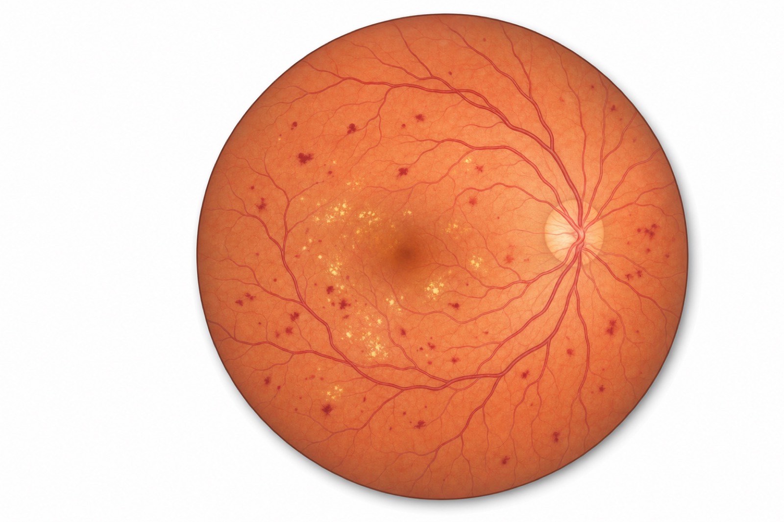
If you have diabetes, your eyes are quietly on the front line of the disease. Diabetic retinopathy, the damage that high blood sugar does to the blood vessels of the retina, is the leading cause of new blindness in working-age adults in the United States, and it is largely preventable when it is caught at the right time. The problem is timing. Diabetic retinopathy can advance for years without any noticeable change in vision, which is exactly why so many people first learn they have it only after it has become serious. The single most effective thing you can do is keep the screening interval most patients miss.
Palm Beach County carries a diabetes burden above the national average, according to data tracked in the CDC Diabetes Atlas, and many of our South Florida patients go far too long between dilated diabetic eye exams. This guide explains what diabetic retinopathy is, the four-stage path it follows, the screening schedule that catches it in time, the imaging we use to find it, and the treatment ladder a retina specialist actually climbs, from blood sugar control to anti-VEGF injections to surgery. It is written for an educated patient and is not a substitute for a dilated eye examination.
What Is Diabetic Retinopathy?
Diabetic retinopathy is damage to the tiny blood vessels of the retina caused by chronically elevated blood sugar. The retina is the light-sensitive tissue at the back of the eye that captures images and sends them to the brain, and it depends on a dense network of small vessels to stay healthy. Over time, diabetes injures those vessels, and the retina pays the price.
How high blood sugar damages retinal blood vessels
Persistently high blood glucose weakens the walls of the small retinal vessels. They can begin to leak fluid and blood, develop tiny bulges called microaneurysms, and eventually become blocked. When vessels close off, areas of the retina are starved of oxygen. The retina responds to that oxygen shortage by signaling for new blood vessels to grow, but the new vessels it builds are abnormal, fragile, and prone to bleeding. This cascade, from leaking to blockage to abnormal new vessel growth, is the core story of diabetic retinopathy and the reason it grows more dangerous as it advances.
Why it is the leading cause of new adult blindness in the US
Diabetic retinopathy is the leading cause of new cases of blindness among working-age adults in the United States, a fact documented by the National Eye Institute. The reason is a combination of how common diabetes is and how silently the eye disease progresses. Because vision often stays normal until late in the disease, many people do not seek care until permanent damage has occurred. The encouraging counterpoint is that the great majority of severe vision loss from diabetic retinopathy is preventable with timely screening and treatment.
Why Most Patients Don't Notice It Until It Is Advanced
The most dangerous feature of diabetic retinopathy is that it does not hurt and, for a long time, does not blur your vision. In the early and even moderate stages, the changes are happening at the level of microscopic blood vessels that do not yet affect the central seeing part of the retina. You can have meaningful, progressing disease and still read the bottom line of an eye chart.
By the time vision does change, the disease has often reached a stage where treatment is more involved and some damage may be permanent. This is the central paradox of diabetic eye disease: the window when treatment is easiest and most effective is precisely the window when you feel completely fine. That is why screening, not symptoms, is the foundation of protecting sight in diabetes. Waiting for blurry vision is waiting too long.
The Four Stages of Diabetic Retinopathy
Diabetic retinopathy is classified into four stages that describe a continuum of worsening vessel damage. The first three are grouped as non-proliferative, meaning no abnormal new vessels have grown yet. The fourth, proliferative, is defined by that abnormal new vessel growth.
Mild non-proliferative (NPDR)
This earliest stage is marked by a small number of microaneurysms, the tiny balloon-like swellings in the retinal vessel walls. Vision is normal, and most patients have no idea anything is wrong. Mild NPDR is usually managed by tightening blood sugar control and monitoring, because catching the disease here, before it progresses, is the goal of screening.
Moderate NPDR
As the disease advances, more microaneurysms appear, small hemorrhages develop, and some retinal vessels begin to close. The retina is starting to lose part of its blood supply. Patients still typically see well, but the risk of progression rises, and the monitoring interval is shortened so that any further change is caught quickly.
Severe NPDR
In severe non-proliferative disease, widespread vessel blockage starves large areas of the retina of oxygen. This is a critical stage, because an oxygen-starved retina is on the verge of triggering abnormal new vessel growth. A substantial fraction of patients with severe NPDR progress to the proliferative stage within a year if nothing changes, so this is a point at which a retina specialist watches very closely and considers whether to begin treatment.
Proliferative diabetic retinopathy (PDR)
Proliferative diabetic retinopathy is the most advanced and sight-threatening stage. In response to oxygen starvation, the retina grows new, abnormal blood vessels (a process called neovascularization). These fragile vessels can bleed into the vitreous gel that fills the eye, causing sudden floaters or vision loss, and they can form scar tissue that pulls on the retina and causes a tractional retinal detachment. PDR can cause severe, permanent vision loss, but it is also highly treatable when addressed promptly with the therapies described below.
Diabetic Macular Edema: A Separate but Related Threat
Diabetic macular edema, or DME, is the most common reason people with diabetes lose vision, and it can occur at any stage of diabetic retinopathy, not just the advanced stages. The macula is the small central part of the retina responsible for sharp, detailed vision, the vision you use to read, recognize faces, and drive. In DME, leaking retinal vessels allow fluid to accumulate in the macula, causing it to swell.
That swelling distorts and blurs central vision. Unlike the rest of diabetic retinopathy, DME can affect your sight relatively early, because it strikes the one part of the retina where even a small amount of fluid is noticeable. The important point is that DME is treatable, often very effectively, with the same anti-VEGF injection therapy used for advanced disease. Because DME can develop even in milder retinopathy, it is one more reason regular dilated exams and retinal imaging matter regardless of how well you think you are seeing.
How Often You Should Get a Dilated Diabetic Eye Exam
The screening interval is the single most important number in this article, and it differs depending on your type of diabetes and your situation. These intervals reflect the recommendations in the AAO Preferred Practice Pattern on Diabetic Retinopathy and the American Diabetes Association Standards of Care.
Type 1 diabetes screening interval
Adults with type 1 diabetes should have their first dilated eye exam within five years of diagnosis, and then at least once a year. The five-year window reflects the fact that retinopathy takes time to develop after type 1 onset, but once that window passes, annual screening is the standard.
Type 2 diabetes screening interval
Because type 2 diabetes is often present for years before it is diagnosed, retinopathy can already exist at the moment of diagnosis. For that reason, people with type 2 diabetes should have a dilated eye exam at the time of diagnosis and then at least once a year thereafter. If exams are consistently normal and blood sugar is well controlled, your ophthalmologist may extend the interval, but only your eye doctor should make that call.
Pregnancy and diabetes
Pregnancy can accelerate diabetic retinopathy, so women with pre-existing type 1 or type 2 diabetes should have a dilated eye exam before pregnancy or in the first trimester, with follow-up during pregnancy and for a year afterward as advised. (This applies to diabetes present before pregnancy; gestational diabetes that develops during pregnancy carries a different, lower risk profile.) If you have diabetes and are pregnant or planning to become pregnant, tell your retina specialist so the right schedule can be set.
What we look for in a dilated retinal exam
In a dilated exam, drops widen your pupils so we can examine the entire retina, not just the center. We look for microaneurysms, hemorrhages, signs of vessel closure, abnormal new vessels, and any swelling of the macula. Dilation is essential because much of the early damage occurs in the periphery of the retina, where it cannot be seen through an undilated pupil. The exam is painless, though your vision will be blurry and light-sensitive for several hours afterward, so it is wise to arrange a ride.
Imaging Used at West Boca Eye Center
A dilated exam is the foundation, but modern retinal imaging lets us detect and measure diabetic changes with far greater precision. We use several complementary technologies.
OCT (optical coherence tomography)
Optical coherence tomography is a quick, non-invasive scan that produces a detailed cross-section of the retina, almost like a CT scan of the macula. OCT is the workhorse for detecting and measuring diabetic macular edema, because it shows fluid and swelling within the retinal layers with great accuracy. It is also how we track whether treatment is working, by measuring how the swelling responds over time.
Fluorescein angiography
Fluorescein angiography involves injecting a harmless dye into a vein in the arm and then photographing the retina as the dye flows through its vessels. This highlights areas where vessels are leaking, blocked, or growing abnormally, giving us a map of the disease that a standard exam cannot. It is particularly useful for identifying areas of poor blood supply and guiding laser treatment.
Ultra-widefield retinal imaging
Traditional retinal photographs capture only the central portion of the retina. Ultra-widefield imaging captures a much larger view in a single image, revealing peripheral damage that might otherwise be missed. Because diabetic retinopathy often begins and progresses in the periphery, this wider view helps us stage the disease accurately and catch advancing changes earlier.
Treatment Options by Stage
Treatment for diabetic retinopathy is matched to the stage and to whether macular edema is present. It climbs as a ladder, from the foundation of systemic control to injections, laser, and finally surgery for the most advanced cases.
Blood sugar, blood pressure, and lipid control (the foundation)
No eye treatment substitutes for controlling the underlying disease. Tight control of blood sugar, blood pressure, and cholesterol is the foundation that everything else is built on, and it is the most powerful tool for slowing or preventing diabetic retinopathy. Landmark diabetes trials have shown that better glucose control meaningfully reduces the risk of retinopathy progression. This is a partnership between you, your primary care physician or endocrinologist, and your retina specialist. The injections and lasers described below treat the damage; good control prevents more of it.
Anti-VEGF injections for diabetic macular edema and PDR
Anti-VEGF therapy is now a first-line treatment for diabetic macular edema and an important treatment for proliferative disease. The abnormal vessel growth and leakage in diabetic retinopathy are driven by a protein called vascular endothelial growth factor, or VEGF. Anti-VEGF medications, which include aflibercept, ranibizumab, and bevacizumab, are injected into the eye to block that signal, reducing macular swelling and causing abnormal vessels to regress. The injection is performed in the office after the eye is numbed and takes only seconds.
The choice of agent and schedule is individualized. The DRCR Retina Network's Protocol T study directly compared these anti-VEGF drugs for diabetic macular edema and helps guide which agent may be preferred in a given situation, but we do not declare a single drug universally "best," because the right choice depends on the individual eye and how it responds. Treatment usually begins as a series of monthly injections and is then extended as the swelling improves.
Focal and panretinal laser photocoagulation
Laser treatment has two distinct roles in diabetic retinopathy. Focal (or grid) laser is used to seal specific leaking vessels contributing to macular edema, often in combination with injections. Panretinal photocoagulation, or PRP, is used for proliferative disease: the laser treats the oxygen-starved peripheral retina to reduce its demand for new vessels, causing the dangerous neovascularization to regress. PRP has been a cornerstone treatment for advanced diabetic retinopathy for decades and remains highly effective at preventing severe vision loss, though it is sometimes used alongside anti-VEGF therapy rather than instead of it.
Vitrectomy surgery for advanced bleeding or traction
When proliferative disease leads to a vitreous hemorrhage that does not clear, or to scar tissue pulling the retina into a tractional detachment, surgery may be needed. Vitrectomy is an operating-room procedure in which the blood-filled vitreous gel is removed, scar tissue is carefully peeled away, and the retina is repaired and stabilized. Vitrectomy is reserved for the most advanced cases, but for those patients it can restore vision that would otherwise be lost.
Symptoms That Should Prompt a Same-Week Call
Because diabetic retinopathy is usually silent, any new visual symptom in a person with diabetes deserves prompt attention. Call your retina specialist the same week, and sooner if symptoms are dramatic, if you experience any of the following:
- A sudden increase in floaters, especially a shower of new spots, which can signal bleeding inside the eye.
- Sudden blurring or loss of vision in one or both eyes.
- A dark area, shadow, or curtain across part of your vision, which can indicate a retinal detachment.
- New distortion, where straight lines look wavy or bent, which can signal macular swelling.
- Flashes of light accompanying new floaters.
These symptoms do not always mean a catastrophe, but in a person with diabetes they should never be ignored or watched for weeks. The same-week phone call is one of the most important habits a diabetic patient can build.
Diabetic Eye Care at West Boca Eye Center
At West Boca Eye Center, diabetic eye care is led by board-certified ophthalmologists with retina expertise, using the full range of imaging and treatment under one roof. We coordinate with your primary care physician and endocrinologist so that your eye care and your overall diabetes management work together. For patients who are due, or overdue, for screening, we make it straightforward to get a thorough dilated exam with OCT and widefield imaging.
In our Boca Raton practice, our goal is to catch diabetic changes at a stage where they can be monitored or treated early, long before vision is threatened. When treatment is needed, we offer anti-VEGF injections, focal and panretinal laser, and vitrectomy surgery for advanced disease. You can learn more about our team on Dr. Nezgoda's bio and Dr. Pagano's bio, and read more about our diabetic retinopathy program and the broader retinal services in Boca Raton we provide. Because diabetic retinopathy is one of several conditions that affect the back of the eye, we also evaluate related concerns such as retinal detachment warning signs and macular degeneration, which also affects the macula. A thorough comprehensive eye exam is the entry point for anyone with diabetes who is unsure when they were last screened.
Important Safety Information
This article is for general education and is not medical advice. Decisions about diabetic eye screening intervals and treatment must be individualized by a qualified ophthalmologist or retina specialist. Anti-VEGF injections (which include aflibercept, ranibizumab, and bevacizumab) and laser photocoagulation are effective treatments but carry risks. Intravitreal injection risks include, but are not limited to, eye infection (endophthalmitis), retinal detachment, increased eye pressure, intraocular inflammation, and, rarely, serious vision loss. Panretinal laser can reduce peripheral and night vision and may affect color vision. Vitrectomy is a surgical procedure with its own risks, including bleeding, infection, retinal detachment, and cataract formation. No single anti-VEGF drug is universally superior; the appropriate agent depends on the individual eye and clinical judgment. Controlling blood sugar, blood pressure, and cholesterol is essential and complements, but does not replace, eye treatment. If you have diabetes and experience new floaters, flashes, sudden blurred or lost vision, or a shadow across your vision, contact your retina specialist the same week, and sooner if symptoms are severe.
Frequently Asked Questions
How often should a diabetic get an eye exam?
People with type 1 diabetes should have a dilated eye exam within five years of diagnosis and then at least annually. People with type 2 diabetes should be examined at diagnosis and then at least annually, because retinopathy may already be present when type 2 is diagnosed. Women with pre-existing diabetes who become pregnant need more frequent monitoring. Your ophthalmologist may adjust the interval based on your findings and control.
Can diabetic retinopathy be reversed?
Early diabetic retinopathy can sometimes stabilize or partially improve with excellent blood sugar, blood pressure, and cholesterol control, but established damage to retinal vessels is generally not fully reversible. The realistic goal is to catch the disease early, slow or halt its progression, and treat sight-threatening complications such as macular edema and proliferative disease before vision is lost. This is why screening before symptoms appear is so important.
Does diabetic retinopathy have symptoms?
Usually not in its early and moderate stages. Diabetic retinopathy is typically painless and does not blur vision until it is advanced or until macular edema develops. This silent course is precisely why regular dilated exams, rather than waiting for symptoms, are the foundation of protecting your sight when you have diabetes.
What are anti-VEGF injections, and do they hurt?
Anti-VEGF injections deliver medication into the eye to reduce macular swelling and shrink abnormal blood vessels in diabetic retinopathy. The eye is thoroughly numbed first, and the injection itself takes only seconds. Most patients are surprised by how tolerable it is. Mild irritation or a red spot on the white of the eye afterward is common and temporary.
What is diabetic macular edema?
Diabetic macular edema is swelling of the macula, the central part of the retina, caused by fluid leaking from damaged retinal vessels. It is the most common cause of vision loss in diabetes and can occur at any stage of diabetic retinopathy. It blurs and distorts central vision but is highly treatable, most often with anti-VEGF injections and sometimes focal laser.
Can I lose my vision from diabetes even if I see fine now?
Yes, which is the central danger of diabetic eye disease. You can have significant, progressing diabetic retinopathy while still seeing clearly, because early damage occurs in parts of the retina that do not yet affect central vision. By the time vision changes, the disease is often advanced. Regular dilated exams catch the problem while it is still treatable and silent.
Will laser treatment for diabetic retinopathy affect my vision?
Panretinal laser treatment for proliferative disease is highly effective at preventing severe vision loss, but it works by treating the peripheral retina, which can reduce some peripheral vision and night vision. This is a worthwhile trade to prevent the far greater vision loss that untreated proliferative disease can cause. Focal laser for macular edema is more targeted. Your retina specialist will explain what to expect for your specific situation.
Schedule a Diabetic Eye Exam in Boca Raton
West Boca Eye Center serves patients from Boca Raton, Delray Beach, Boynton Beach, Deerfield Beach, Parkland, and Coral Springs. If you have diabetes and cannot remember your last dilated eye exam, or if you have noticed any change in your vision, do not wait. Call our office this week to schedule a diabetic eye exam, or request an appointment through our contact page. Catching diabetic retinopathy early is one of the most reliable ways to protect your sight for the long term.
Book an appointment
Fill out the form below and our staff will reach out to you quickly to fully book your appointment and receive all of your necessary information.

Specializing in modern cataract surgery.
Located 1/2 miles North of West Boca Medical Center on Glades Road, directly behind Macy's Furniture Gallery.
West Boca Eye Center
9325 Glades Road, Suite 201.
Boca Raton, FL 33434