


Post Category


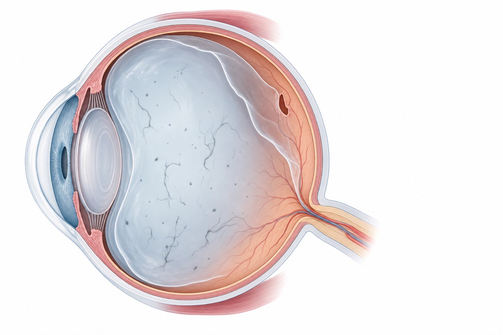
Almost everyone develops floaters eventually. Those little specks, threads, and cobwebs that drift across your vision are usually harmless, and in South Florida we see them constantly in patients in their 50s and 60s. But here is the part that matters: the very same symptoms, new floaters and flashes of light, can also be the first warning sign of a retinal tear or detachment, a true emergency that can steal sight within hours to days if it is not treated. The symptoms can look identical to you. Only an exam can tell them apart. So before anything else, let us start with triage.
Triage First: When New Floaters Are an Emergency
If you take only one thing from this article, take this. The following symptoms mean you should be seen urgently, the same day or the next day, by an ophthalmologist for a dilated exam. Do not wait to see whether they improve. Call our office immediately if you notice any of these.
Sudden shower of new floaters
A few floaters that drift in slowly over weeks are usually benign. A sudden burst of many new floaters all at once, sometimes described as a swarm of gnats or a spray of black dots or soot, is different. It can indicate that the gel inside the eye has pulled on or torn the retina, or that there is bleeding inside the eye. A sudden shower of new floaters is a same-day or next-day exam, not a wait-and-see.
Flashes of light, especially in the peripheral vision
Flashes that look like brief streaks, arcs, or lightning bolts, particularly off to the side of your vision, are a signal that the retina is being tugged. They are caused by traction on the retina, and they often accompany the separation of the vitreous gel from the retinal surface. New flashes, especially together with new floaters, warrant an urgent dilated exam to make sure the retina has not torn.
A dark curtain or shadow across part of your vision
This is the most urgent symptom of all. A dark curtain, veil, or shadow advancing across any part of your field of vision can mean the retina is actively detaching. A retinal detachment is a true emergency, and the amount of vision that can be saved depends directly on how quickly it is treated. If you see a curtain or shadow coming across your vision, this is a right-now problem. Seek care immediately.
Recent eye trauma or post-cataract-surgery floaters
The risk calculation changes if you have specific risk factors. New floaters or flashes after a blow to the eye or head, after recent cataract surgery, or in someone who is highly nearsighted, carry a higher likelihood of a retinal tear and should be evaluated promptly. The same is true for anyone with a personal or family history of retinal detachment. If any of these apply to you, do not wait on new symptoms; call right away.
What Floaters Actually Are
Now that the urgent signs are covered, here is what is actually happening in the eye, because understanding the mechanism helps explain why most floaters are harmless and why a few are not.
The vitreous gel and why it casts shadows
The large cavity in the back of the eye is filled with a clear, jelly-like substance called the vitreous. When you are young, this gel is firm and uniform. With age, it gradually becomes more liquid and develops small clumps and strands of collagen. When light enters the eye, these clumps cast tiny shadows on the retina, and those shadows are what you perceive as floaters. According to the National Eye Institute, floaters are extremely common and usually a normal part of aging.
Why floaters look like specks, threads, or cobwebs
Because floaters are actually shadows of structures suspended in moving gel, they appear to drift when you move your eyes and then settle when your eye stops. They take many shapes: dots, circles, threads, squiggles, or cobweb-like strands. They tend to be most noticeable against a bright, plain background, such as a clear blue sky or a white wall or screen. A floater that has been present and stable for months is almost always benign. It is the new, the sudden, and the accompanied-by-flashes floaters that need attention.
Posterior Vitreous Detachment (PVD): The Most Common Cause
The single most common reason for a sudden onset of new floaters and flashes is a posterior vitreous detachment, or PVD. This is not the same as a retinal detachment, and the names are unfortunately easy to confuse.
Why it happens with age, typically 50 to 70
As the vitreous gel liquefies with age, it eventually shrinks and pulls away from the retina at the back of the eye. This separation is a PVD. It is a normal, age-related event that most people experience, typically between the ages of 50 and 70. When the gel separates, it can tug on the retina (producing flashes) and a clump where it was attached can become newly visible (producing a prominent floater, sometimes ring-shaped). For our Boca Raton patients in this age range, a PVD is one of the most common reasons for a sudden floater.
Why higher myopia patients get PVD earlier
People who are significantly nearsighted (high myopia) have longer eyes, and the vitreous in a longer eye tends to liquefy and separate earlier in life. As a result, highly myopic patients often experience PVD years sooner than average and also carry a somewhat higher lifetime risk of retinal tears. If you have always worn strong glasses or contacts for distance, your eyes deserve extra vigilance when new floaters appear.
How long PVD floaters stick around
Once a PVD has occurred and the retina has been confirmed intact, the floaters it produces usually become less bothersome over weeks to months. The brain learns to ignore them, the floater may drift out of the central line of sight, and the flashes typically fade as the traction resolves. Most people adapt well and do not require any treatment. The key qualifier is the phrase confirmed intact, which is why even a typical PVD warrants a dilated exam.
Retinal Tears and Detachments: The Threat Hiding Inside a PVD
Here is the reason a PVD is not something to simply shrug off. As the vitreous pulls away, it can occasionally pull hard enough to tear the retina. A retinal tear can then allow fluid to seep underneath the retina and lift it off, which is a retinal detachment.
Why up to 1 in 7 acute PVD patients have a retinal tear
This is the statistic every patient with new floaters should know. A systematic review by Hollands and colleagues, published in JAMA in 2009, found that among patients who present with acute, symptomatic floaters and flashes, roughly 1 in 7 has a retinal tear at the initial examination. That is why the symptoms cannot be safely self-diagnosed. The majority of people with acute floaters have a benign PVD, but a meaningful minority have a tear that needs treatment, and there is no way to tell which group you are in without a dilated look at the retina.
What an in-office dilated exam rules in or out
The exam that settles the question is a dilated fundus examination. Drops widen the pupil, and the ophthalmologist examines the entire retina, including the far periphery where tears usually occur, often with gentle pressure on the eye to view the very edges. This exam can definitively identify or rule out a retinal tear or detachment. It is painless, takes only a short time, and is the single most important step after new floaters or flashes appear. If the retina is intact, you can be reassured. If a tear is found, it can usually be treated the same day.
How retinal tears are treated (laser retinopexy or cryotherapy)
A retinal tear caught before it progresses to a detachment can often be sealed in the office, which is exactly why early evaluation matters so much. Laser retinopexy uses a laser to create tiny spot welds around the tear, sealing the retina down so fluid cannot get under it. Cryotherapy (freezing treatment) achieves a similar seal in certain locations. Both are office procedures designed to prevent a tear from becoming a detachment. If a detachment has already occurred, more involved surgery is generally required, which is why sealing a tear early can save a patient from a far larger operation and protect vision.
Other Causes of Floaters
While PVD is the most common cause, new floaters can occasionally arise from other conditions, some of which are also urgent.
Vitreous hemorrhage
Bleeding into the vitreous, called a vitreous hemorrhage, produces a sudden shower of floaters, often described as smoke, soot, or a red haze, and can cloud vision significantly. It can result from a torn retinal blood vessel, advanced diabetic eye disease, or other causes. A vitreous hemorrhage always warrants prompt evaluation to find the source.
Uveitis
Uveitis is inflammation inside the eye. It can release inflammatory cells and debris into the vitreous that are perceived as floaters, usually accompanied by eye redness, pain, and light sensitivity. Uveitis requires medical treatment of the underlying inflammation and should be evaluated by an ophthalmologist.
Diabetic eye disease
In people with diabetes, abnormal, fragile blood vessels can grow on the retina and bleed into the vitreous, producing new floaters. Any patient with diabetes who develops new floaters should be evaluated promptly, because it can signal advancing diabetic retinopathy that needs treatment.
When Is It Safe to 'Watch and Wait'?
So when is a floater truly nothing to worry about? In general, a single floater, or a small number of floaters, that has been present and unchanged for months, with no flashes, no curtain or shadow, and no sudden increase, is very likely a benign, long-standing finding. Stable floaters that your brain has already learned to ignore do not require urgent attention.
The crucial caveat is that watch and wait is only appropriate after the retina has been confirmed healthy, and for symptoms that are old and stable, not new. Any new floater, any flashes, any change in an existing floater, or any of the red-flag symptoms at the top of this article moves you out of the watch-and-wait category and into the get-examined category. When in doubt, the safest course is always a dilated exam. An exam that turns out reassuring is never a wasted visit.
Treatment Options for Persistent Floaters
Most floaters need no treatment beyond the reassurance of a normal exam and time for the brain to adapt. For the minority of patients whose floaters remain genuinely disabling, a few options exist, and candidacy matters.
Observation
Observation is the first-line approach for almost all floaters. After a PVD with a confirmed intact retina, the great majority of patients find their floaters fade in prominence and stop interfering with daily life within weeks to months. No procedure carries zero risk, so for most people the right choice is to let the eye settle and the brain adapt.
YAG laser vitreolysis (laser floater removal)
YAG laser vitreolysis is an in-office laser procedure that targets and breaks up certain floaters to make them less noticeable. It is not right for everyone. The American Academy of Ophthalmology notes that laser vitreolysis is selective: it tends to work best for specific types of floaters, such as a single, well-defined, centrally located clump (like the ring-shaped floater that can follow a PVD), and is less suitable for diffuse, cloud-like, or near-the-retina floaters. According to the American Academy of Ophthalmology, careful patient selection is essential. A thorough evaluation determines whether you are a candidate for our laser floater removal at WBEC.
Vitrectomy
Vitrectomy is a surgical procedure that removes the vitreous gel, and with it the floaters, replacing it with a clear solution. It is the most definitive treatment but also the most invasive, with surgical risks that include retinal detachment, infection, and cataract formation. For that reason it is reserved for severe, persistent, vision-impairing floaters in carefully selected patients after other options have been considered. Whether vitrectomy is appropriate is a decision made individually with a retina specialist.
What to Expect at a Same-Day WBEC Visit
If you call us with new floaters or flashes, our goal is to see you quickly, the same day or the next day, for a dilated examination. When you arrive, we will check your vision and eye pressure, then place dilating drops, which take about 20 to 30 minutes to widen your pupils fully. Once dilated, the ophthalmologist examines the entire retina, including the periphery, to look for any tear, detachment, hemorrhage, or other cause of your symptoms.
If the retina is intact, we will reassure you and explain what to watch for going forward. If we find a retinal tear, it can often be treated the same day with laser or cryotherapy to seal it before it progresses. Because your eyes will be dilated and your vision blurry and light-sensitive for several hours, please arrange for someone to drive you home, and bring sunglasses. The visit is straightforward, and the peace of mind from a definitive answer is well worth it.
Important Safety Information
This article is for general education and is not medical advice. New or sudden floaters, new flashes of light, or a shadow or curtain across your vision can signal a retinal tear or detachment and require prompt evaluation by an ophthalmologist, typically a same-day or next-day dilated exam. Do not attempt to self-diagnose the cause of new floaters; only a dilated retinal examination can reliably distinguish a benign posterior vitreous detachment from a retinal tear. Among patients with acute symptomatic floaters and flashes, roughly 1 in 7 has a retinal tear at presentation, as reported by Hollands and colleagues in JAMA. Treatments for floaters carry risks: YAG laser vitreolysis is appropriate only for selected floaters and candidates and can, uncommonly, cause complications including elevated eye pressure, lens damage, or retinal injury; vitrectomy is a surgical procedure with risks including retinal detachment, infection, bleeding, and cataract formation. The decision to treat, and the choice of procedure, must be individualized by a qualified ophthalmologist. If you experience a sudden shower of floaters, flashes of light, or a curtain across your vision, seek care immediately.
Frequently Asked Questions
Are eye floaters dangerous?
Most floaters are harmless, especially those that have been stable for months. However, a sudden increase in floaters, new floaters with flashes of light, or a shadow across your vision can signal a retinal tear or detachment, which is an emergency. Because you cannot reliably tell a benign floater from a dangerous one on your own, any new or sudden floater should be checked with a dilated eye exam.
When should I worry about floaters and flashes?
Seek urgent care, the same day or next day, if you experience a sudden shower of many new floaters, new flashes of light (especially in your side vision), or a dark curtain or shadow moving across your field of view. These can be signs of a retinal tear or detachment. The risk is higher if you are very nearsighted, had recent eye trauma or cataract surgery, or have a history of retinal detachment.
What is a posterior vitreous detachment?
A posterior vitreous detachment, or PVD, is the age-related separation of the eye's vitreous gel from the retina, most common between ages 50 and 70. It is the most frequent cause of sudden new floaters and flashes and is usually benign. However, the same pulling action that causes a PVD can occasionally tear the retina, which is why a PVD should be confirmed safe with a dilated exam.
How likely is a retinal tear if I have new floaters and flashes?
According to a systematic review published in JAMA by Hollands and colleagues, roughly 1 in 7 patients who present with acute, symptomatic floaters and flashes has a retinal tear at the first examination. Most people have a benign posterior vitreous detachment instead, but because a meaningful minority have a tear, a dilated exam is essential to tell them apart.
Can floaters go away on their own?
Often, yes, in the sense that they become less noticeable. After a benign posterior vitreous detachment with a confirmed healthy retina, most floaters fade in prominence over weeks to months as they drift out of the line of sight and the brain learns to ignore them. The floater material does not usually disappear entirely, but it typically stops interfering with daily life. Treatment is reserved for floaters that remain genuinely disabling.
Is laser floater removal right for me?
It depends on the type of floater and your eye. YAG laser vitreolysis works best for certain well-defined, centrally located floaters and is not appropriate for diffuse floaters or those close to the retina. It is not suitable for everyone, and careful patient selection is essential. The only way to know if you are a candidate is a thorough evaluation with an ophthalmologist who offers the procedure.
What happens during a dilated eye exam for floaters?
We first check your vision and eye pressure, then place dilating drops that take 20 to 30 minutes to widen your pupils. The ophthalmologist then examines your entire retina, including the periphery, to look for any tear, detachment, or bleeding. If the retina is intact, you are reassured. If a tear is found, it can often be sealed the same day with laser or freezing treatment. Your vision will be blurry for several hours, so arrange a ride and bring sunglasses.
Call West Boca Eye Center If You Have New Floaters or Flashes
West Boca Eye Center serves patients from Boca Raton, Delray Beach, Boynton Beach, Deerfield Beach, Parkland, and Coral Springs. If you have a sudden shower of new floaters, new flashes of light, or any shadow across your vision, do not wait. Call our office right away for a same-day or next-day dilated exam, or reach us through our contact page. For symptoms that may be an emergency, we also provide emergency eye care. You can read more about our retinal care program, retinal detachment in detail, and retinal holes and tears, and learn about our team on Dr. Pagano's bio and Dr. Nezgoda's bio pages.
Book an appointment
Fill out the form below and our staff will reach out to you quickly to fully book your appointment and receive all of your necessary information.

Specializing in modern cataract surgery.
Located 1/2 miles North of West Boca Medical Center on Glades Road, directly behind Macy's Furniture Gallery.
West Boca Eye Center
9325 Glades Road, Suite 201.
Boca Raton, FL 33434