
Post Category


If you live in Boca Raton, spend your weekends on the water, or simply commute under the year-round South Florida sun, there is a good chance you have seen a pterygium without knowing its name. A pterygium (pronounced tuh-RIJ-ee-um) is a fleshy, wedge-shaped growth of tissue that creeps from the white of the eye onto the clear cornea, usually on the side closest to the nose. Patients often call it "surfer's eye," because chronic sun, wind, and dust exposure are the main drivers, and surfers, boaters, fishermen, and outdoor workers develop it at higher rates. In a coastal, high-ultraviolet climate like ours, pterygium is one of the most common reasons a patient lands in an ophthalmologist's chair.
This guide explains what a pterygium actually is, how to tell it apart from a pinguecula, when it can simply be watched versus when it needs surgery, and what modern pterygium surgery (conjunctival autograft) involves. It is written for an educated patient by a board-certified ophthalmologist practicing in Boca Raton. It is not a substitute for a comprehensive eye examination.
Why pterygium is so common in South Florida
Pterygium prevalence tracks closely with sun exposure. Populations that live closer to the equator, work outdoors, and spend more time in reflective environments such as water and sand develop pterygium far more often than populations in cooler, cloudier regions. South Florida checks every box.
The UV plus wind plus dust formula
The single most important risk factor is cumulative ultraviolet light exposure, particularly UV-B. According to the American Academy of Ophthalmology, pterygium is strongly associated with a lifetime of sun exposure, which is why it is sometimes grouped with other UV-related ocular surface changes. Wind and airborne dust add chronic mechanical irritation and dryness to the ocular surface, and that combination of UV plus drying plus micro-irritation is what drives the conjunctival tissue to thicken and advance onto the cornea.
Year-round exposure compounds the risk
In much of the country, sun exposure is seasonal. In Boca Raton and across Palm Beach County, it is a year-round reality. There is no winter break from the ultraviolet load. Patients who golf, boat, fish, run, cycle, or work outdoors here are accumulating UV exposure twelve months a year, and that steady, decades-long dose is exactly what a pterygium feeds on. It is also why we counsel patients so heavily on protection: the same sun that caused the growth in the first place is the sun that drives it to come back after surgery.
Pterygium vs pinguecula: what's the difference
These two are frequently confused, and they are related, but they are not the same thing.
- A pinguecula is a yellowish, slightly raised deposit on the conjunctiva (the white of the eye), usually on the nasal side. It sits on the white of the eye and does not grow onto the cornea. It rarely affects vision.
- A pterygium is a fibrovascular growth that extends from the conjunctiva onto the cornea (the clear front window of the eye). Because it can encroach on the cornea, it can affect vision when it grows far enough or distorts the corneal shape.
A pinguecula can sometimes progress into a pterygium over years of continued exposure, but many never do. The practical distinction is simple: a pinguecula stays on the white, a pterygium crosses onto the clear cornea. You can learn more about how we evaluate both on our pinguecula and pterygium service page.
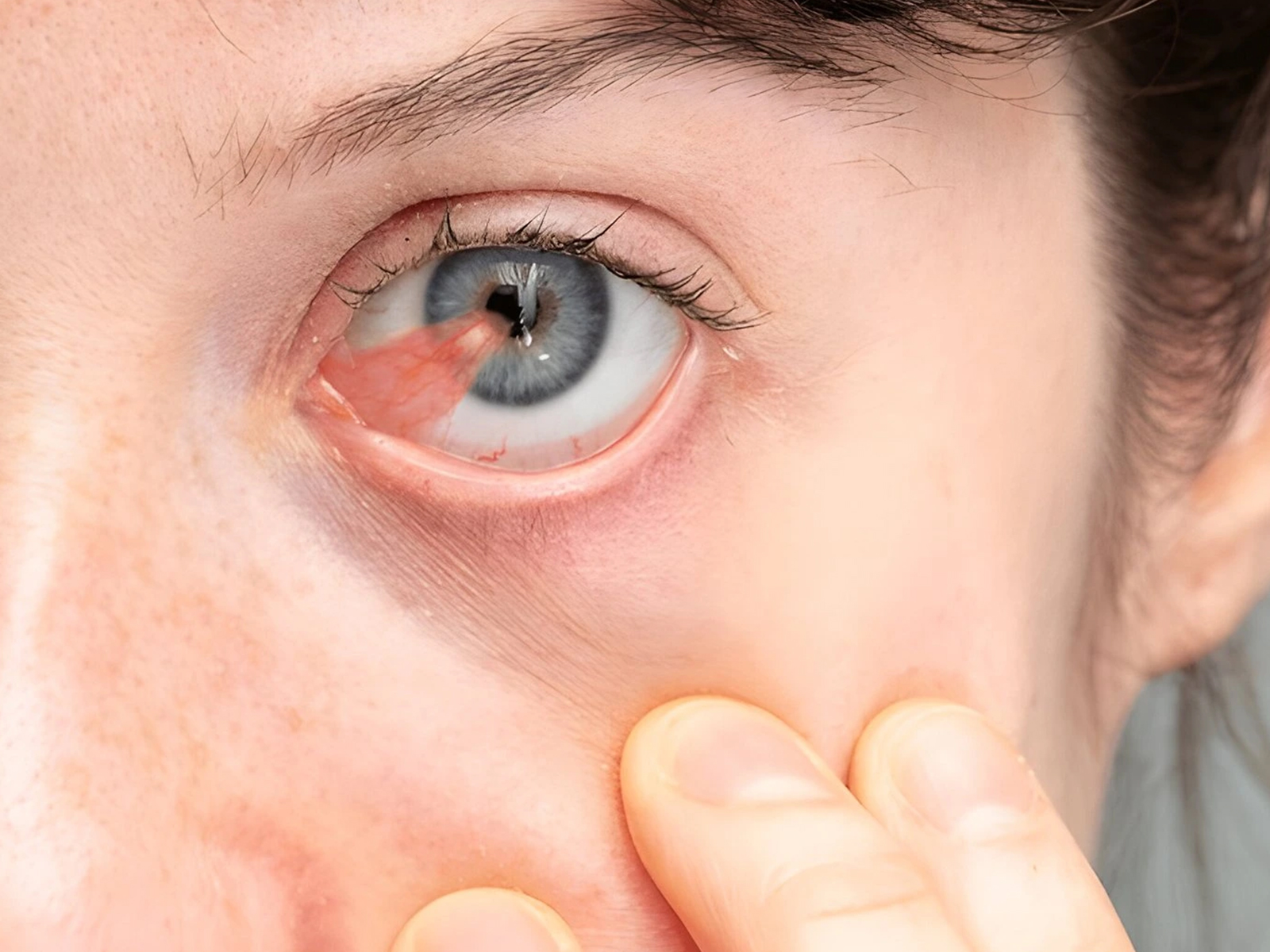
What pterygium actually is
Anatomy: a fibrovascular growth of conjunctiva onto cornea
A pterygium is a wing-shaped fold of conjunctival tissue, rich in blood vessels and fibrous tissue, that grows across the limbus (the border between the white of the eye and the cornea) and onto the cornea itself. The word pterygium comes from the Greek for "little wing," which describes its triangular shape. Most pterygia point toward the center of the cornea from the nasal side, although they can occur on the temporal (outer) side as well.
Why it's not cancer
This is the first question most patients ask, and the answer is reassuring: a pterygium is a benign (non-cancerous) growth. It is a reaction of the ocular surface to chronic environmental stress, not a tumor. That said, any growth on the eye that changes in appearance, grows rapidly, or has unusual pigmentation should be evaluated by an ophthalmologist, because rare ocular surface lesions can mimic a pterygium. A proper examination distinguishes a benign pterygium from a lesion that needs different attention.
How it can affect vision
A small pterygium often causes no visual symptoms at all. As it advances, it can affect vision in two ways. First, as the tissue pulls on the corneal surface, it can induce astigmatism, distorting the cornea and blurring vision even before the growth reaches the visual axis. Second, if the pterygium grows far enough across the cornea to encroach on the central visual axis, it can directly block clear vision. Either situation is a reason to consider surgical removal rather than continued observation.
Symptoms most Boca Raton patients notice first
Redness and irritation
The most common early complaint is a persistently red or bloodshot-looking area on the inner corner of the eye, especially after a day in the sun, wind, or salt air. The blood vessels within the pterygium become engorged and inflamed, and the eye can look chronically irritated.
A visible wedge near the nose-side of the eye
Many patients notice a raised, fleshy, triangular patch of tissue on the white of the eye, closest to the nose, that seems to be slowly creeping toward the colored part of the eye. Cosmetic concern is a legitimate and common reason patients seek evaluation.
Foreign body sensation
Because the pterygium is a raised growth, it disrupts the smooth tear film across the eye. That often produces a gritty, sandy, or "something in my eye" sensation, along with intermittent tearing. The disruption of the tear film also overlaps with dry eye, which is extremely common in our air-conditioned, screen-heavy South Florida environment. For patients whose primary complaint is dryness and irritation, we often evaluate for both conditions together; you can read more on our dry eye syndrome page.
Blurry or fluctuating vision
When a pterygium induces astigmatism or approaches the visual axis, patients notice blur that may fluctuate during the day, particularly when the eye is dry or irritated. This is one of the clearest signals that a pterygium has progressed from a cosmetic and comfort issue to a functional one.
When to watch vs when to treat
Not every pterygium needs surgery. In fact, most do not, at least not right away. The decision to observe versus operate depends on symptoms, growth, and effect on vision.
Conservative management
For a small, stable, asymptomatic or mildly symptomatic pterygium, conservative management is usually the right first step:
- Lubrication. Preservative-free artificial tears reduce the gritty sensation and calm surface irritation. A lubricating gel at night can help patients who wake with dryness.
- UV protection. Consistent use of wraparound, UV-blocking sunglasses and a wide-brim hat reduces the ongoing stimulus that drives growth. This is the single most important conservative measure.
- Short-term anti-inflammatory drops. During flare-ups of redness and swelling, your ophthalmologist may prescribe a brief course of mild anti-inflammatory or vasoconstrictor drops. These are used short-term and under supervision, not as a daily habit, because chronic use of certain drops can cause its own problems.
Conservative care does not make a pterygium disappear. It controls symptoms and slows progression while we monitor the growth over time.
When surgery becomes the right answer
We typically move toward surgical removal when one or more of the following is true:
- The pterygium is encroaching on or threatening the visual axis.
- It is inducing significant astigmatism and degrading vision.
- Symptoms of irritation, redness, and foreign body sensation are persistent and not controlled by conservative measures.
- The growth is documented to be advancing across the cornea.
- The patient has a significant cosmetic concern that affects quality of life.
- The pterygium interferes with contact lens wear.
Surgery is the only way to actually remove a pterygium. The goal is to remove it cleanly and, just as importantly, to reduce the chance that it comes back.
Modern pterygium surgery: conjunctival autograft
Why bare-sclera excision is no longer standard
For decades, the standard technique was "bare-sclera" excision: the surgeon simply removed the pterygium and left the underlying white of the eye (sclera) exposed to heal on its own. The problem was recurrence. Bare-sclera excision has historically been associated with high recurrence rates reported in the literature, in some series approaching or exceeding fifty percent, which is why it has largely been abandoned as a stand-alone technique. When a pterygium recurs, it often comes back more aggressively and with more scarring than the original.
The conjunctival autograft technique
The modern standard of care is excision with a conjunctival autograft. After the pterygium is removed, the surgeon takes a small, thin piece of healthy conjunctiva from elsewhere on the same eye (typically from under the upper eyelid, where it is hidden) and transplants it to cover the bare area left behind. Because the graft comes from your own eye, there is no rejection risk. This technique dramatically lowers recurrence compared with bare-sclera excision and produces a healthier, smoother ocular surface. Peer-reviewed cornea literature consistently shows conjunctival autografting to be the most effective approach for reducing recurrence, which is the central goal of pterygium surgery.
Fibrin glue vs sutures
The graft has to be secured in place. There are two common ways to do this:
- Sutures. Fine, dissolvable or removable stitches hold the graft. Sutures are reliable but can cause more post-operative irritation and a longer period of foreign body sensation while they are present.
- Fibrin tissue glue. An FDA-cleared fibrin sealant (a biological "glue" derived from clotting proteins) can be used to bond the graft in place without stitches. Many surgeons and patients prefer glue because it is associated with less post-operative discomfort and shorter operating time. As with any blood-derived product, your surgeon will discuss the specifics with you.
The choice between sutures and glue is surgeon-dependent and is individualized to your eye and the size of the graft.
Adjunctive mitomycin C: when it's used
In certain higher-risk or recurrent cases, surgeons may use a brief intraoperative application of mitomycin C, a medication that reduces the regrowth of abnormal tissue. Its use is adjunctive and surgeon-dependent. It is not used in every case, because it carries its own risk profile, and the decision is made selectively based on factors such as recurrence history and the appearance of the tissue. This is a conversation to have with your surgeon, not a default add-on.
Recovery and what to expect
The first week
Pterygium surgery is an outpatient procedure performed under local anesthesia, usually in well under an hour. You go home the same day. Expect the eye to be red, watery, and to feel scratchy or irritated for the first several days, particularly if sutures were used. We prescribe anti-inflammatory and antibiotic drops, and most patients are comfortable with over-the-counter pain relief. The graft will look red and noticeable at first; this is expected and improves steadily.
Weeks two through six
Redness and irritation taper over the following weeks. The graft gradually blends in and the surface smooths out. Most patients return to normal daily activities within a week or two, while strenuous exercise, swimming, and any activity that exposes the eye to dust, wind, or water are restricted longer per your surgeon's instructions. Follow-up visits let us confirm the graft is healing and watch for any early signs of recurrence, which is most likely in the first several months to a year.
Long-term care
Long-term, the most important job is protection. The eye is healed, but the environment that caused the original pterygium has not changed. Lifelong, consistent UV protection is what keeps a recurrence from forming.
Recurrent pterygium: prevention is everything
Why recurrence happens
Recurrence is the central challenge of pterygium surgery. The growth is fundamentally a response to environmental stress, so if the stimulus continues unchecked, the tissue can be driven to grow back. Recurrence risk is highest in younger patients, in those with more aggressive original growths, and in those who return to heavy unprotected sun exposure. This is exactly why technique matters so much: the conjunctival autograft exists to lower this risk in the first place.
The role of post-op UV protection
The behavioral half of recurrence prevention belongs to the patient. Wraparound UV-blocking sunglasses, a wide-brim hat, and consistent lubrication after surgery are not optional extras in South Florida; they are part of the treatment. We are blunt about this with our Boca Raton patients, because a beautiful surgical result can still recur if the eye goes back to year-round unprotected sun.
What to do if it comes back
If a pterygium recurs, do not panic, but do come in for evaluation. Recurrent pterygia can be managed surgically again, often with a conjunctival autograft (or an amniotic membrane graft) and, in select cases, adjunctive mitomycin C. The approach is individualized, because recurrent tissue tends to be more scarred and more vascular than the original.
Preventing pterygium in Boca Raton's climate
UV-blocking sunglasses with wraparound coverage
Standard sunglasses block light from the front but let UV in around the sides, where the nasal conjunctiva is most exposed. Wraparound styles that block 100 percent of UV-A and UV-B provide far better protection for the exact area where pterygium forms. This is the highest-value prevention step you can take.
Wide-brim hats and lubricating drops
A wide-brim hat adds another layer of shade, and preservative-free artificial tears keep the ocular surface healthy and less reactive to wind and dust. Together with sunglasses, these are simple, inexpensive, and effective.
Why year-round protection matters here
The defining feature of pterygium prevention in South Florida is that it never goes off-season. The sun in January is still strong, the reflection off the water and sand is constant, and outdoor life here is a twelve-month affair. Patients who protect their eyes only on "beach days" still accumulate substantial exposure on every ordinary sunny day in between. Year-round protection is the rule that actually keeps pterygium from forming or returning.
Important Safety Information
Pterygium surgery is a safe and effective procedure in appropriately selected patients, but no eye surgery is risk-free. Risks of pterygium excision with conjunctival autograft include recurrence, infection, inflammation, graft displacement or loss, bleeding, scarring, persistent redness or foreign body sensation, induced astigmatism, and, rarely, loss of best-corrected vision. Adjunctive mitomycin C, when used, carries additional risks and is reserved for selected cases at the surgeon's discretion. Conservative management does not eliminate a pterygium and is not a cure. Any new, rapidly growing, or unusually pigmented growth on the eye should be evaluated promptly, because rare ocular surface lesions can resemble a pterygium. Candidacy for pterygium surgery requires a comprehensive evaluation with a board-certified ophthalmologist.
Frequently Asked Questions
Is a pterygium dangerous?
A pterygium itself is a benign, non-cancerous growth and is not dangerous in the way a tumor would be. It can, however, affect vision if it grows onto the cornea, induces astigmatism, or reaches the visual axis. Any growth that changes rapidly or looks unusual should be examined to rule out other, rarer conditions.
Will a pterygium go away on its own?
No. A pterygium does not resolve on its own. Conservative measures such as lubrication and UV protection can control symptoms and slow progression, but the only way to remove a pterygium is surgically.
What is the difference between a pterygium and a pinguecula?
A pinguecula is a yellowish bump on the white of the eye that does not grow onto the cornea. A pterygium is a fibrovascular growth that extends from the white of the eye onto the clear cornea. A pinguecula can sometimes progress into a pterygium over time, but many never do.
Does pterygium surgery hurt?
The surgery itself is performed under local anesthesia, so you should not feel pain during the procedure. Afterward, the eye is typically red, watery, and scratchy for several days, particularly if sutures were used. Most patients manage discomfort with prescribed drops and over-the-counter pain relief.
How likely is a pterygium to come back after surgery?
Recurrence risk depends heavily on the technique used and on post-operative sun protection. The older bare-sclera technique had high recurrence rates, which is why the conjunctival autograft is now the standard of care, since it substantially lowers recurrence. Consistent year-round UV protection after surgery further reduces the risk.
Can I prevent a pterygium if I live in South Florida?
You can significantly reduce your risk. Wraparound UV-blocking sunglasses, a wide-brim hat, and lubricating drops protect the ocular surface from the UV, wind, and dust that drive pterygium formation. In a year-round sun climate, consistent protection is the key.
Will removing a pterygium improve my vision?
If the pterygium was inducing astigmatism or encroaching on the visual axis, removal can improve vision. If it had not yet affected vision, surgery is performed primarily for comfort, surface health, or cosmetic reasons rather than for visual improvement.
How long is recovery after pterygium surgery?
Most patients return to normal daily activities within one to two weeks, with redness and irritation tapering over several weeks. Strenuous activity, swimming, and exposure to wind, dust, and water are restricted longer per your surgeon's instructions. Follow-up visits monitor healing and watch for early recurrence.
Get your pterygium evaluated in Boca Raton
West Boca Eye Center serves patients from Boca Raton, Delray Beach, Boynton Beach, Deerfield Beach, Parkland, and Coral Springs. If you have a growth on the white of your eye, persistent redness and irritation, or vision that has begun to blur, a comprehensive evaluation can tell you whether you are dealing with a pinguecula, a pterygium, or something else, and whether observation or surgery is the right next step. To schedule with Dr. Brent Bellotte, a board-certified Boca Raton ophthalmologist, visit our Boca Raton location page or book a comprehensive eye exam.
Pterygium is common in sunny coastal Florida. A Boca Raton guide to what causes it, when to treat it, and what modern pterygium surgery actually involves.
Book an appointment
Fill out the form below and our staff will reach out to you quickly to fully book your appointment and receive all of your necessary information.
Specializing in modern cataract surgery.
Located 1/2 miles North of West Boca Medical Center on Glades Road, directly behind Macy's Furniture Gallery.
West Boca Eye Center
9325 Glades Road, Suite 201.
Boca Raton, FL 33434